| Home | E-Submission | Sitemap | Contact Us |  
Journal of Neurocritical Care > Volume 16(1); 2023 > Article
Kim, Sung, Lee, and Park: Posterior reversible encephalopathy syndrome superimposed on neuronal intranuclear inclusion disease
A 54-year-old female patient presented to the emergency room with vomiting and convulsions, which subsided after the administration of 4 mg of intravenous (IV) lorazepam. The patient was previously diagnosed with neuronal intranuclear inclusion disease (NIID) at another tertiary hospital. The patient was stuporous and had a fever (39.2 °C) and elevated blood pressure (180/90 mm Hg).
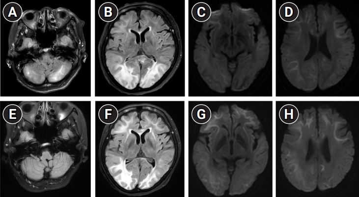
Brain magnetic resonance imaging (MRI) showed high signal intensity in the bilateral parietooccipital lobe and cerebellum on fluid-attenuated inversion recovery (FLAIR), which was suggestive of posterior reversible encephalopathy syndrome (PRES) (Fig. 1A and B) [1,2]. Additionally, MRI revealed bilateral diffusion restriction in the corticomedullary junction on diffusion-weighted imaging (DWI), in accordance with NIID (Fig. 1C and D) [3,4].
Her blood pressure was meticulously adjusted using IV nicardipine [2]. The patient slowly improved and experienced drowsiness. Follow-up brain MRI revealed less prominent FLAIR high signal intensity, which implied improved PRES (Fig. 1E and F). However, the MRI findings of NIID were aggravated, and lesions were more prominent in the bilateral frontotemporal lobes (Fig. 1G and H). At discharge, the patient was alert, and her cognitive impairment and apraxia persisted [4].
Neurologists should always consider the possibility of other conditions, such as PRES, when managing patients with NIID with drowsy mental status and excessively high blood pressure. The highly variable symptoms of NIID might confuse physicians [3].

ARTICLE INFORMATION

Ethics statement
This study was reviewed and approved by the Institutional Review Board of Hanyang University Hospital (No. 2022-10-013). The need for informed consent from the patient was waived by the board.
Conflict of interest
No potential conflict of interest relevant to this article.
Author contributions
Conceptualization: ES. Methodology: NK. Formal analysis: NK.Data curation: NK. Visualization: ES. Project administration: NK. Funding acquisition: NK. Writing–original draft: NK. Writing–review & editing: SL, JP.

Fig. 1.
Brain magnetic resonance imaging scans performed 12 hours after the seizure (A-D) and after 3 weeks of treatment (E, F). (A, B) Fluid-attenuated inversion recovery (FLAIR) revealed high signal intensity lesion at the bilateral parietooccipital lobe, right temporal lobe, and cerebellum. (E, F) The follow-up image showed less prominent high FLAIR signal intensity at the cerebellum and parietooccipital lobes. (C, D) Diffusion-weighted imaging (DWI) showed bilateral symmetric diffusion restriction lesions in the corticomedullary junction, at the frontal, parietal, and temporal lobes. (G, H) The follow-up DWI showed slightly worsened diffusion restriction at the bilateral frontotemporal lobes.
jnc-220084f1.jpg

REFERENCES

1. Triplett JD, Kutlubaev MA, Kermode AG, Hardy T. Posterior reversible encephalopathy syndrome (PRES): diagnosis and management. Pract Neurol 2022;22:183-9.
crossref pmid
2. Fugate JE, Rabinstein AA. Posterior reversible encephalopathy syndrome: clinical and radiological manifestations, pathophysiology, and outstanding questions. Lancet Neurol 2015;14:914-25.
crossref pmid
3. Sone J, Mori K, Inagaki T, Katsumata R, Takagi S, Yokoi S, et al. Clinicopathological features of adult-onset neuronal intranuclear inclusion disease. Brain 2016;139(Pt 12):3170-86.
crossref pmid pmc
4. Chi X, Li M, Huang T, Tong K, Xing H, Chen J. Neuronal intranuclear inclusion disease with mental abnormality: a case report. BMC Neurol 2020;20:356.
crossref pmid pmc pdf
Editorial Office
Department of Neurology and Critical Care, Seoul National University Hospital,
101, Daehak-ro, Jongno-gu, Seoul, 03080 Korea
TEL: +82-2-2072-0743 (10:00 AM ~ 4:00 PM) / +82-2-2072-1810   FAX: +82-2-3672-7553    E-mail: office@e-jnc.org

Copyright© Korean Neurocritical Care Society.                Developed in M2PI