급성 간부전에서의 두개내압 상승의 병태생리와 치료

Pathophysiology and Treatment of Cerebral Edema in Acute Liver Failure

Article information

J Neurocrit Care. 2016;9(2):63-70
Publication date (electronic) : 2016 December 9
doi : https://doi.org/10.18700/jnc.160088
1Division of Gastroenterology and Hepatology, Department of Internal Medicine, Kyungpook National University School of Medicine, Daegu, Korea
2Department of Neurology, Gyeongsang National University Changwon Hospital, Changwon, Korea
장세영1, 장준영,2
1경북대학교 의과대학ㆍ의학전문대학원 내과학교실 소화기내과
2창원경상대학교병원 신경과
Corresponding Author: Jun Young Chang  Department of Neurology, Gyeongsang National University Changwon Hospital, 11 Samjeongja-ro, Seongsan-gu, Changwon 51472, Korea  Tel: +82-55-214-3813 Fax: +82-55-214-2638 E-mail: kjychang@gmail.com
Received 2016 October 31; Revised 2016 November 11; Accepted 2016 November 21.

Trans Abstract

Early diagnosis and management of cerebral edema in acute liver failure is important to reduce neurological complication and mortality. Ammonia-induced astrocyte swelling and increased blood brain barrier permeability via transmembrane dysfunction are major mechanisms of cerebral edema in acute liver failure. Conventional therapy can be used to lower intracranial pressure. In addition, various treatment options are available to reduce serum ammonia level. Herein, we described the pathophysiology, monitoring, and management of cerebral edema in acute liver failure.

서 론

두개골이라는 한정된 공간에 뇌척수액(10%, 140 mL), 혈액(10%, 140 mL) 및 뇌실질(80%, 1,120 mL)이 포함되어 있으며 각각의 구성 성분이 조화를 이루어 두개내압을 일정하게 유지한다[1]. 여러 가지 원인으로 인해 보상 기전과 균형이 깨어지게 되면 두개내압이 상승하게 되는데, 세 가지 구성요소 중 가장 많은 비중을 차지하고 있는 뇌실질의 부종이 가장 흔한 두개내압 상승의 원인이다[2]. 뇌부종에 의한 두개내압 상승은 뇌질환 이외에도 급성 간부전으로 인한 간성 뇌병증, 콩팥 기능 저하로 인한 투석 환자에서의 투석 불균형 증후군, 고혈압성 뇌병증 등의 전신 질환과 동반되어서도 나타날 수 있으며 조기에 진단해서 치료하는 것이 중요하다. 본 논문에서는 뇌부종을 초래할 수 있는 전신 질환 중 간질환으로 인한 뇌부종의 원인 및 병태 생리와 치료 방침에 관해 알아보고자 한다.

본 론

1. 급성 간부전과 간성 뇌병증

급성 간부전(acute liver failure)은 기저 간질환이 없는 환자에서 갑자기 발생하는 심한 간세포의 소실로 인한 임상 증후군으로 사망률이 80%에 달하며 이러한 사망 원인 중 일부는 심한 뇌부종으로 인한 두개내압 상승과 뇌 탈출에 기인한다. 급성 간부전의 발생 원인으로 아세트아미노펜과 비아세트아미노펜에 의한 약물 유발 간독성이 50% 이상을 차지하며 그 외 B형 간염(7%), 다른 바이러스에 의한 간염(3%), 자가면역 간염(5%), 허혈성 간염(4%) 등이 있다[3]. 간성 뇌병증은 황달의 발생에서부터 뇌병증이 발생하는 기간에 따라 1주일 이내의 초급성기, 8일에서 28일 사이의 급성기, 5-12주 사이의 아급성기로 분류하기도 하며 초급성으로 빠르게 뇌병증으로 진행하는 경우에 뇌부종이 발생할 위험성이 가장 높다[4,5].

간성 뇌병증의 경우 의식 수준 및 인지기능 정도에 따라 단계를 구분하게 되며 침상에서도 평가가 가능한 West Haven criteria를 많이 사용한다(Table 1) [6,7]. 3단계 환자의 25-35%, 4단계 환자의 65~75% 에서 급성 간부전으로 인한 뇌부종이 발생하며 암모니아의 배출 장애로 인한 축적이 중요한 원인으로 알려져 있다[8,9]. 별아교세포(astrocyte)의 팽윤으로 인한 세포독성 부종과 혈액뇌장벽의 손상으로 인한 혈관성 부종 두 가지 기전이 모두 급성 간부전 환자에서의 뇌부종 발생에 영향을 미친다.

Severity of hepatic encephalopathy (West Haven criteria)

2. 별아교세포의 팽윤으로 인한 세포독성 부종

지속적으로 세포 내로 유입되는 나트륨, 칼슘과 같은 양이온 및 글루탐산염 (glutamate)과 같은 신경전달 물질을 세포막에 존재하는 아데노신 삼인산염 (adenosine triphosphate, ATP) 의존성 Na+/K+ 이온통로를 포함한 여러 이온 채널들이 세포 밖으로 배출하여 세포 내외의 이온 균형을 유지하게 된다. 허혈이나 저산소 손상 등 다양한 원인에 의해 뇌세포가 손상을 받게 되면 세포의 기능을 유지하게 위해 필수적인 ATP의 생성이 줄어들게 된다. ATP가 부족하게 되면 나트륨, 칼슘 등의 양이온이 세포 밖으로 배출되지 못한 채 지속적으로 내부에 축적되고, 이온 균형을 맞추기 위해 염소 이온과 같은 음이온이 유입되어 세포 내부의 삼투압이 증가하게 된다[10,11]. 증가된 세포 내부의 삼투압으로 인해 Aquaporin 통로를 통해 수분이 유입되어 세포 내의 부피가 증가하고 수포 형태의 표면 변화가 발생하면서 부풀어 오르다 사멸하는 것이 세포독성 부종이다[11,12].

급성 간부전에서는 세포독성 부종의 발생에 암모니아가 주된 역할을 하고 글루탐산염, 염증성 사이토카인, 젖산, 괴사된 간으로부터 유리되는 물질, 고온, 저나트륨혈증 등이 추가적인 상승효과를를 일으키는 것으로 알려져 있다[13-15]. 특히 이러한 세포독성 부종은 뇌전체 부피의 1/3을 차지하며 회색질에 분포하고 있는 별아교세포의 발돌기(foot process)에 주로 발생한다[16]. 암모니아는 소장에 있는 글루탐산 분해 효소(glutaminase)에 의해 글루타민이 글루탐산염과 암모니아로 분해되면서 생성되며 이러한 암모니아의 대부분이 간의 요소회로 (urea cycle)을 통해 대사된다. 급성 간기능 손상으로 인해 혈중 암모니아가 분해되지 못하고 축적되게 되면 확산을 통해 혈액뇌장벽을 통과한 후 별아교세포 내로 유입되어 세포 부종을 초래하게 된다. 기존에는 암모니아가 별아교세포 내부에서 글루타민으로 재합성되어 축적된 글루타민이 삼투물질로 작용하여 세포 부종을 초래하는 것으로 알고 있었으나 글루타민의 농도와 세포 부종의 정도 및 시간적 선후 관계가 잘 맞지 않는다는 보고가 있어[17,18], 최근에는 별아교세포 내부에서 재합성된 글루타민이 미토콘드리아로 유입되면서 phosphate-activated glutaminase (PAG)에 의해 암모니아와 글루탐산염으로 다시 분해되고 이로 인한 산화 스트레스가 세포 부종을 유발한다는 설명이 유력하다(Trojan Horse hypothesis, Fig. 1) [19-21].
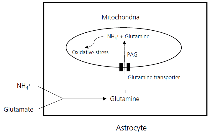
Figure 1.

Cytotoxic astrocyte swelling due to oxidative stress in the mitochondria. The excess glutamine transported into mitochondria and degraded into ammonia and glutamate by phosphate-activated glutaminase (PAG). Ammonia and glutamate accumulated inside the mitochondria produce reactive oxygen species, resulting in astrocyte swelling.

3. 혈관 뇌 장벽의 손상으로 인한 혈관성 부종

과거 연구에서는 급성 간부전으로 인한 뇌부종의 경우 혈액 뇌장벽의 구조에는 큰 변화가 없어 주로 세포독성 부종에 의한 현상으로 파악하였으나, 최근 연구에서는 치밀이음부(tight junction)를 구성하는 단백질의 미세한 기능 변화와 전신 염증성 사이토카인의 영향으로 인한 혈액뇌장벽 투과도의 변화로 인해 혈관성 부종이 함께 발생하는 것으로 밝혀졌다.

혈액뇌장벽은 모세혈관 내피세포 및 치밀이음부, 혈관주위 세포(pericyte), 별아교세포의 발돌기로 이루어져 있으며 이중 내피 세포와 치밀이음부가 장벽으로서의 기능을 하는 데 중요한 역할을 한다. 특히 내피세포간 치밀이음부에 존재하는 occludin, claudin-5, junctional adhesion molecule (JAM), cadherin과 같은 여러 가지의 막경유단백들이 세포주변 통로(paracellular path)를 통한 물질의 이동에 관여한다.

급성 간부전을 유도한 쥐의 혈액뇌장벽을 전자 현미경으로 관찰하였을 때 치밀이음부의 구조적인 변화는 명확하지 않으나 여러 다양한 염색 물질들의 투과도는 증가하는 현상을 관찰하였고 이는 구조적인 변화보다는 치밀이음부에 존재하는 막경유단백질의 기능 변화로 인한 현상으로 판단하고 있다. 특히 급성 간부전시에 괴사한 간세포로부터 혈관 내로 유리되는 tumor necrosis factor alpha, interleukin-1, interleukin-6 등 염증성 사이토카인이 Toll-like receptor 4/NF-kB 경로를 활성화 시켜 치밀이음부 단백의 발현을 억제하고 Matrix metalloproteinase-9 (MMP-9)이 치밀이음부에 존재하는 occludin, claudin-5와 같은 단백질을 분해함으로써 혈액뇌장벽의 투과도를 증가시킨다[22-24]. 또한 암모니아가 MMP-9의 활성 및 활성산소 (reactive oxygen species), 산화 질소(nitric oxide)의 생성을 증가시켜 혈액뇌장벽의 투과도를 증가시키기도 한다[25].

4. 뇌부종 발생 유무에 대한 평가 및 모니터

급성 간부전 환자에서 뇌부종 발생을 예측하는 위험 인자로는 3단계 이상의 간성 뇌병증이 발생한 경우, 혈중 암모니아 수치가 100 umol/L를 초과하는 경우, 초급성으로 간성 뇌병증이 발생하는 경우, 전신성 염증반응 증후군(sysmetic inflammatory response syndrome)이 동반된 경우, 신대치 요법이나 승압제가 필요한 경우 등이 있다[19,26]. 급성 간부전으로 인한 간성 뇌병증 환자에서 두통, 구토, 의식 상태 악화 등과 같은 임상 증상 만으로는 뇌부종 발생 여부를 조기에 진단하기에 불충분하다. 혈압이 갑자기 상승하면서 서맥 및 불규칙한 호흡을 보이는 쿠싱 반사, 동공 반사 및 전정안구 반사(oculovestibular reflex)의 이상, 대뇌 제거 자세(decerebrate posture) 역시 뇌부종이 진행된 상태에서 나타나므로 조기 진단에 활용하기에는 적절하지 않다. 고위험 환자에서 시기 적절한 뇌부종의 진단과 치료를 위해 뇌 컴퓨터단층촬영(CT), 뇌 자기공명 영상(MRI), 뇌혈류 초음파(TCD)와 같은 뇌영상 검사가 필요하다.

뇌 CT를 통해 혈액응고장애로 인해 발생할 수 있는 뇌출혈을 배제할 수 있고 뇌고랑 소실, 뇌백질과 회색질의 경계 소실, 뇌바닥 수조의 압박, 수두증을 관찰함으로써 뇌부종 발생 유무를 평가할 수 있다[27]. 뇌 MRI의 확산강조영상을 통해 뇌부종의 발생 위치와 정도를 좀 더 민감하게 확인할 수 있고 겉보기확산계수(apparent diffusion coefficient) 영상을 통해 세포 독성 부종과 혈관성 부종을 감별할 수 있으나 검사에 시간이 오래 걸리고 장시간 바로 누운 자세를 유지해야 하는 위험이 있다[3]. 뇌혈류 초음파는 뇌혈관의 자동 조절 능력이 손상된 상황에서는 뇌혈류량이 뇌압과 비례한다는 사실을 근거로 뇌혈류 속도를 측정해서 간접적으로 뇌압의 변화 양상을 비침습적으로 모니터 할 수 있다. 뇌부종이 발생하게 되면 가장 먼저 중대뇌 동맥의 수축기 혈류 속도가 증가하다가 점차 뇌압이 상승하면서 혈류 속도는 감소하게 되고 박동지수(pulsatility index) 는 증가하게 된다. 최대 수축기 혈류 파형이 날카로워지고 뇌내 혈관의 탄성도가 감소하면서 혈관의 탄성반동(elastic recoil)이 소실됨으로 인해 수축기 때의 혈류 파형이 변화하게 된다(loss of the Windkessel notch, Fig. 2) [28].

Figure 2.

Transcranial doppler waveform of middle cerebral artery from a patient with acute liver failure. Under normal circumstances, the first peak is related to myocardial contractility and the second peak is produced by the distensibility of the arterial wall (asterisk indicates diastolic vascular recoil). As intracranial pressure increases, peak systolic velocity increases and wave becomes sharpened due to external compression of the arter

간이식이 필요한 3, 4단계 간성 뇌병증 환자 및 아세트아미노펜에 의한 급성 간부전과 같이 간이식의 적응증은 아니나 추후 회복될 여지가 있는 환자군에서는 정확한 두개내압 측정과 변동 양상에 대한 빠른 평가를 위해 두개내압측정 카테터를 이용한 모니터가 필요하며 뇌실질내에 직접 삽입하는 방법이 가장 정확하다[29]. 응고병증이 동반되어 있는 경우 INR 1.5 미만을 유지하기 위해 신선냉동혈장 (fresh frozen plasma)을 먼저 투여하고 이후에도 교정이 되지 않을 경우에는 재조합 활성 제 8인자(recombinant factor VIIIa)의 투여를 고려해 보아야 한다[30]. 혈소판 감소증이 동반되어 경우 혈소판 수를 100,000/μL 이상 유지하기 위해 예방적인 혈소판 수혈 후에 시술을 고려해야 출혈의 위험성을 줄일 수 있다[31]. 혈청내 피브리노겐(fibrinogen)이 100 mg/dL 미만인 경우에는 동결침전제제 (cryoprecipitate)를 투여한 이후에 시술을 고려해 볼 수 있다. 목정맥팽대 카테터를 이용한 정맥내 산소포화도 측정 (jugular bulb oximetry, SjvO2 >80% indicates hyperemia, SjvO2<50% indicates insufficient oxygen supply), Licox 카테터를 이용한 뇌조직 산소 농도 측정 (Pbto2 <10 mmHg indicates tissue hypoxia), microdialysis를 이용한 뇌조직내 에너지 대사 측정(lactate/pyruvate ratio >40 indicates metabolic crisis)이 두개내압 상승 환자를 집중 관리하는 데 도움이 될 수 있으나 대부분의 연구가 외상성 뇌손상 환자를 대상으로 되어 있어 간부전에 의한 뇌부종 환자에게 적용하는 데에는 근거가 부족한 상태이다[27].

5. 급성 간부전 환자의 내과적 치료

급성 간부전 환자에게는 여러 장기 손상이 동반될 수가 있는데 심장 기능 및 부신 기능 저하로 인한 혈역학적 불안정 및 조직 저관류, 폐기능 저하로 인한 저산소혈증, 콩팥 기능 저하로 인한 복강내 부종 및 위장관벽의 부종, 면역 기능 저하로 인한 감염증, 응고 장애로 인한 출혈 등이 발생할 수 있다[32]. 따라서 급성 간부전 환자의 치료는 환자의 대사와 혈역학적 안정을 유지하여 간의 재생을 돕고 합병증을 최소화하는 것이 근간이 되겠다. 급성간부전에서는 저혈당이 호발하므로 당을 혈액내로 주입하는 것이 필요하지만 저나트륨혈증과 뇌부종을 악화시킬 수 있어 저장성 용액을 대량 주입하는 것은 피해야 한다. 또한, 급성 간부전 상태에서는 에너지가 다량 소모되고 단백질 이화작용(protein catabolism)이 되므로 적절한 영양을 공급하여 환자의 근육양과 면역 기능이 유지되도록 해야 한다. 간성 뇌병증이 있는 경우에는 경구로 1.0-1.5 g/kg/day 가량의 단백질을 공급하고 혈중 암모니아 수치를 모니터링하면서 양이 과다하지 않게 조절한다[33]. 간이식은 급성 간부전 환자의 생존율을 개선시킬 수 있는 확실한 치료법이긴 하나 일반적으로 환자의 병세가 매우 급격하게 진행하고 공여간의 확보가 쉽지 않아 급성 간부전 환자의 약 10%에서만 이루어 진다고 알려져 있다[34,35]. 특히, 뇌사자 공여 보다는 생체 부분 간이식을 시행할 수밖에 없는 경우가 많아 환자가 급성 간부전으로 진단되는 즉시 공여자를 준비하고 환자 상태가 악화되었을 때 응급으로 간이식을 시행할 수 있도록 하는 것이 필요하다. 이번 단락에서는 급성 간부전 환자에서 발생할 수 있는 여러 합병증 중 뇌부종과 이로 인한 두개내압 상승에 대한 치료 방침에 대해서 자세히 살펴보고자 한다.

6. 급성 간부전에서 발생하는 뇌부종의 치료

1) 일반적인 치료

뇌부종 조절의 일반적인 목표는 뇌내압 20 mmHg 미만, 뇌관류압 60 mmHg 이상을 유지하는 것이며 다른 원인으로 인한 두개내압 상승과 비교하여 기본적인 치료 방법에는 큰 차이가 없다. 간성 뇌병증과 두개내압 상승으로 인해 의식이 저하된 환자에서 조기에 빠른 기관 삽관을 하고 기계 환기를 적용하여 저산소혈증을 예방하고 이산화탄소의 농도를 적절히 유지해야 한다 (PCO2 35–40 mmHg) [36]. 적절한 수준의 진정(Ramsay sedation scale 3–5)과 통증 조절, 뇌관류압이 적절히 유지되는 범위 내에서 머리를 30도 세워두는 자세를 유지함으로써 두개내압을 낮춰줄 수 있다[36]. 그 외 발열 및 고혈당 조절(목표 혈당 140–180 mg/dL), 저나트륨혈증에 대한 교정, 락툴로즈 관장을 통해 혈중 암모니아 농도를 떨어뜨리는 치료가 두개내압 상승을 줄여주는데 도움이 된다[27].

2) 약물 치료

삼투압 요법으로 가장 많이 쓰이는 약물은 만니톨과 고장 식염수이다. 만니톨은 0.25–2.0 g/kg의 용량으로 6시간 간격으로 정맥내로 투여한다. 기존에 알려진 혈중 삼투압 농도(serum osmolarity)의 절대값보다는 삼투압 농도차(osmolar gap <55 mOsm/L)를 모니터링 하면서 투여량과 기간을 조절하는 것이 추천되고 있고 적절한 수분 공급을 통해 만니톨 투여중에 발생할 수 있는 혈량 저하증을 예방하는 것이 중요하다[36-38]. 고장성 식염수는 만니톨에 비해 혈액뇌장벽을 비교적 덜 통과하면서 좀 더 강력한 삼투 효과를 낼 수 있다는 장점이 있어 최근 두개내압을 낮추는 약물 치료로 많이 사용되고 있다. 국내에는 11.7% 염화나트륨 주사액 (40 meq/20 mL) 원액 60 mL를 중심정맥카테터나 말초중심정맥카테터(peripherally inserted central catheter, PICC)를 통해 20분에 걸쳐 서서히 투여한다. 혈중 나트륨 농도 145–155 mmol/L를 목표로 6시간 또는 8시간 간격으로 투여한다. 고장성 식염수는 만니톨과 달리 삼투성 이뇨작용이 없고 혈관내 용적을 증가시킬 수 있어 간경화나 심기능 상실 환자와 같은 용적 과부하 상태에서는 조심스럽게 투여해야 한다[39].

3) 저체온 치료

체온이 낮아지면 1도당 6-10% 가량 뇌의 대사가 감소하면서 에너지 요구량이 줄어들고, 세포막이 안정화되면서 나트륨 이온과 칼슘 이온의 무분별한 유입과 이로 인한 뇌세포의 부종, 미토콘드리아 기능 저하, 단백질 분해, 자유라디칼의 생성으로 인한 흥분성 신경전달 물질의 무분별한 분비가 줄어든다[40,41]. 또한 저체온 치료는 세포자멸사를 억제하고 뇌조직으로의 다형핵 백혈구의 유입과 염증 유발 사이토카인의 분비를 줄여 염증 반응을 감소시킴으로써 허혈이나 외상에 의한 뇌질환에서 신경보호 효과 및 두개내압 감소 효과를 보여준다[40].

급성 간부전으로 인한 뇌부종과 두개내압 상승에서 저체온 치료가 생존율 향상에 도움이 된다는 전향적 연구 자료는 없다. 3단계 이상의 간성 뇌병증 환자들에게 저체온 치료(32–35°C)를 시행한 97명의 환자를 저체온 치료를 시행하지 않은 1,135명의 환자와 후향적으로 비교한 연구에서 저체온 치료가 뇌출혈이나 감염증을 증가시키지는 않았으나 21일 생존율에도 큰 영향을 미치지는 못하였다[42]. 그러나 실험을 통해 저체온 치료가 급성 간부전으로 인한 뇌부종의 개선이 도움이 된다는 보고들이 있다. 저체온시에 장내세균에 의해서 암모니아 생성이 감소되고, 콩팥에서 혈중으로 유리되는 암모니아가 감소하면서 혈중 암모니아 농도가 떨어지게 되고, 동시에 뇌혈류 감소로 인해 혈중에서 뇌안으로의 암모니아 흡수가 줄어들면서 뇌내 암모니아 농도가 감소하게 된다[41,43,44]. 저체온 치료는 급성 간부전에서 간성 뇌병증과 뇌부종을 유발하는 것으로 알려져 있는 뇌내 알라닌과 젖산 농도를 감소시키고 세포외 기질에 글루탐산염이 축적되는 것을 줄여 두개내압을 감소시킨다(Table 2) [45-47].

The effects of hypothermia on reducing intracranial pressure (ICP) in acute liver failure.

저체온 치료시 33–35°C 범위로 목표 체온을 설정하고 가능한 한 조기에 목표 체온에 도달하도록 하며 평균 동맥압(80–110 mmHg)과 중심정맥압(8–12 cmH20)을 목표 범위로 유지하기 위한 지속적인 감시가 필요하다. 저체온 요법 중 흔히 발생하는 떨림은 심혈관계 합병증 발생을 증가시키고 호흡일량을 증가시켜 환자의 예후를 악화시키는 요인으로 이를 방지하기 위해 체온조절 담요를 이용해서 피부를 따뜻하게 유지해 주는 것이 중요하다. 체온 조절 담요로 조절되지 않는 경우 여러 약제들 중 간기능이 저하된 경우에도 비교적 안전하게 사용할 수 있는 MgSO4(4 g 15분간 정맥 일시 투여 후 0.5–1.0 g/hour지속 투여, 혈중 농도 3–4 mg/dL 유지), dexmedetomidine (0.3–1.5 μg/kg/hour 지속 정맥 투여), cisatracurium(3 μg/kg/min 정맥 일시 투여 후 0.5–10 μg/kg/min 지속 투여) 등의 약제들을 사용해 볼 수 있다[48].

4)실험적인 치료법들

급성 간부전을 유발한 동물 모델에서 minocycline의 투여가 oxidative, nitrosative stress를 줄임으로써 간성 혼수와 뇌부종이 악화되는 것을 막아주는 역할을 한다는 보고가 있으며[49], N-methyl-D-aspartate (NMDA) 수용체 길항제인 memantine이 NMDA 수용체를 통해 암모니아 독성이 발현되는 것을 억제함으로써 뇌부종을 줄여 주었다는 연구결과가 있다[50,51]. 그 외 혈장 교환술을 통해 두개내압 상승 없이 뇌관류압과 뇌내 산소 대사율(cerebral metabolic rate for oxygen, CMRO2)을 증가시켜 일부 환자의 의식 상태를 개선시켰으며 면역 시스템과 내피 세포 기능을 호전시켜 간이식이 부적합한 환자의 생존율을 증가시킬 수 있다는 주장도 있다[52]. 아세트아미노펜을 이용해서 급성 간부전을 유도한 돼지 모델에서 내독소(endotoxin)를 제거하고 손상된 알부민을 대체해 주는 체외간기능 보조 기구(extracorporeal liver support device)를 이용해서 암모니아 농도 및 염증성 사이토카인의 감소, 뇌압 감소, 평균 동맥압 증가를 통해 생존율을 증가시켰다는 실험 결과도 보고된 바가 있다[53].

결 론

급성 간부전에서 암모니아와 글루탐산에 의한 별아교세포의 팽윤과 세포 독성 부종, 혈액뇌장벽의 치밀이음부 구조 단백의 미세 기능 변화로 인한 혈관성 부종이 뇌부종 유발하여 두개내압을 상승시킨다. 뇌부종 발생 유무에 대한 모니터 및 치료는 뇌의 허혈 손상이나 재관류 손상, 외상에 의한 뇌부종과 근본적으로는 동일하다. 그러나 다른 질환에 의한 뇌부종과 달리 동반된 다발 장기 부전 및 감염증에 의한 혈역학적 불안정에 대한 교정을 통해 적절한 뇌관류압을 유지하기 위한 노력, 두개내압 상승에 중요한 역할을 하는 뇌내 암모니아의 농도를 낮추기 위한 치료가 중요하다. 또한 기계 환기시 진정을 위한 약제 사용이나 저체온 치료시 떨림 부작용을 줄이기 위한 약제 사용시 간으로 대사 되는 약제 선택에 제한이 있다는 점, 고농도의 나트륨을 이용한 삼투압 치료시 용적 과부하 상태의 악화 가능성에 대한 고려가 필요하다는 차이점이 있다.

References

1. Mokri B. The Monro-Kellie hypothesis: applications in CSF volume depletion. Neurology 2001;56:1746–8.
2. Mayer SA, Coplin WM, Raps EC. Cerebral edema, intracranial pressure, and herniation syndromes. J Stroke Cerebrovasc Dis 1999;8:183–91.
3. Mohsenin V. Assessment and management of cerebral edema and intracranial hypertension in acute liver failure. J Crit Care 2013;28:783–91.
4. Bernal W, Auzinger G, Dhawan A, Wendon J. Acute liver failure. Lancet 2010;376:190–201.
5. O'Grady JG, Schalm SW, Williams R. Acute liver failure: redefining the syndromes. Lancet 1993;342:273–5.
6. Montagnese S, Amodio P, Morgan MY. Methods for diagnosing hepatic encephalopathy in patients with cirrhosis: a multidimensional approach. Metab Brain Dis 2004;19:281–312.
7. Cash WJ, McConville P, McDermott E, McCormick PA, Callender ME, McDougall NI. Current concepts in the assessment and treatment of hepatic encephalopathy. QJM 2010;103:9–16.
8. Munoz SJ. Difficult management problems in fulminant hepatic failure. Semin Liver Dis 1993;13:395–413.
9. Shawcross DL, Wendon JA. The neurological manifestations of acute liver failure. Neurochem Int 2012;60:662–71.
10. White BC, Sullivan JM, DeGracia DJ, O'Neil BJ, Neumar RW, Grossman LI, et al. Brain ischemia and reperfusion: molecular mechanisms of neuronal injury. J Neurol Sci 2000;179:1–33.
11. Liang D, Bhatta S, Gerzanich V, Simard JM. Cytotoxic edema: mechanisms of pathological cell swelling. Neurosurg Focus 2007;22:E2.
12. Amiry-Moghaddam M, Ottersen OP. The molecular basis of water transport in the brain. Nat Rev Neurosci 2003;4:991–1001.
13. Jalan R, Olde Damink SW, Hayes PC, Deutz NE, Lee A. Pathogenesis of intracranial hypertension in acute liver failure: inflammation, ammonia and cerebral blood flow. J Hepatol 2004;41:613–20.
14. Blei AT, Olafsson S, Therrien G, Butterworth RF. Ammoniainduced brain edema and intracranial hypertension in rats after portacaval anastomosis. Hepatology 1994;19:1437–44.
15. Norenberg MD. Astrocytic-ammonia interactions in hepatic encephalopathy. Semin Liver Dis 1996;16:245–53.
16. Scott TR, Kronsten VT, Hughes RD, Shawcross DL. Pathophysiology of cerebral oedema in acute liver failure. World J Gastroenterol 2013;19:9240–55.
17. Zwingmann C, Chatauret N, Rose C, Leibfritz D, Butterworth RF. Selective alterations of brain osmolytes in acute liver failure: protective effect of mild hypothermia. Brain Res 2004;999:118–23.
18. Jayakumar AR1, Rao KV, Murthy ChR, Norenberg MD. Glutamine in the mechanism of ammonia-induced astrocyte swelling. Neurochem Int 2006;48:623–8.
19. Tofteng F, Hauerberg J, Hansen BA, Pedersen CB, Jorgensen L, Larsen FS. Persistent arterial hyperammonemia increases the concentration of glutamine and alanine in the brain and correlates with intracranial pressure in patients with fulminant hepatic failure. J Cereb Blood Flow Metab 2006;26:21–7.
20. Sonnewald U, Westergaard N, Jones P, Taylor A, Bachelard HS, Schousboe A. Metabolism of [U-13C5] glutamine in cultured astrocytes studied by NMR spectroscopy: first evidence of astrocytic pyruvate recycling. J Neurochem 1996;67:2566–72.
21. Albrecht J, Norenberg MD. Glutamine: a Trojan horse in ammonia neurotoxicity. Hepatology 2006;44:788–94.
22. Chen G, Zhang S, Shi J, Ai J, Qi M, Hang C. Simvastatin reduces secondary brain injury caused by cortical contusion in rats: possible involvement of TLR4/NF-kappaB pathway. Exp Neurol 2009;216:398–406.
23. Aslam M, Ahmad N, Srivastava R, Hemmer B. TNF-alpha induced NFkappaB signaling and p65 (RelA) overexpression repress Cldn5 promoter in mouse brain endothelial cells. Cytokine 2012;57:269–75.
24. Chen F, Radisky ES, Das P, Batra J, Hata T, Hori T, et al. TIMP-1 attenuates blood-brain barrier permeability in mice with acute liver failure. J Cereb Blood Flow Metab 2013;33:1041–9.
25. Skowronska M, Zielinska M, Wojcik-Stanaszek L, Ruszkiewicz J, Milatovic D, Aschner M, et al. Ammonia increases paracellular permeability of rat brain endothelial cells by a mechanism encompassing oxidative/nitrosative stress and activation of matrix metalloproteinases. J Neurochem 2012;121:125–34.
26. Bernal W, Hall C, Karvellas CJ, Auzinger G, Sizer E, Wendon J. Arterial ammonia and clinical risk factors for encephalopathy and intracranial hypertension in acute liver failure. Hepatology 2007;46:1844–52.
27. Rabinstein AA. Treatment of brain edema in acute liver failure. Curr Treat Options Neurol 2010;12:129–41.
28. Aggarwal S, Brooks DM, Kang Y, Linden PK, Patzer JF 2nd. Noninvasive monitoring of cerebral perfusion pressure in patients with acute liver failure using transcranial doppler ultrasonography. Liver Transpl 2008;14:1048–57.
29. Stravitz RT, Kramer AH, Davern T, Shaikh AO, Caldwell SH, Mehta RL, et al. Intensive care of patients with acute liver failure: recommendations of the U.S. Acute Liver Failure Study Group. Crit Care Med 2007;35:2498–508.
30. Mahajan A, Lat I. Correction of coagulopathy in the setting of acute liver failure. Crit Care Nurs Clin North Am 2010;22:315–21.
31. Liumbruno GM, Bennardello F, Lattanzio A, Piccoli P, Rossetti G, ; Italian Society of Transfusion Medicine and Immunohaematology Working Party. Recommendations for the transfusion management of patients in the peri-operative period. III. The post-operative period. Blood Transfus 2011;9:320–35.
32. Wang DW, Yin YM, Yao YM. Advances in the management of acute liver failure. World J Gastroenterol 2013;19:7069–77.
33. Bernal W, Wendon J. Acute liver failure. N Engl J Med 2013;369:2525–34.
34. Simpson KJ, Bates CM, Henderson NC, Wigmore SJ, Garden OJ, Lee A, et al. The utilization of liver transplantation in the management of acute liver failure: comparison between acetaminophen and non-acetaminophen etiologies. Liver Transpl 2009;15:600–9.
35. Germani G, Theocharidou E, Adam R, Karam V, Wendon J, O'Grady J, et al. Liver transplantation for acute liver failure in Europe: outcomes over 20 years from the ELTR database. J Hepatol 2012;57:288–96.
36. Jeon SB, Koh Y, Choi HA, Lee K. Critical care for patients with massive ischemic stroke. J Stroke 2014;16:146–60.
37. Torre-Healy A, Marko NF, Weil RJ. Hyperosmolar therapy for intracranial hypertension. Neurocrit Care 2012;17:117–30.
38. Wijdicks EF, Sheth KN, Carter BS, Greer DM, Kasner SE, Kimberly WT, et al. Recommendations for the management of cerebral and cerebellar infarction with swelling: a statement for healthcare professionals from the American Heart Association/American Stroke Association. Stroke 2014;45:1222–38.
39. Torbey MT, Bosel J, Rhoney DH, Rincon F, Staykov D, Amar AP, et al. Evidence-based guidelines for the management of large hemispheric infarction : a statement for health care professionals from the Neurocritical Care Society and the German Society for Neuro-intensive Care and Emergency Medicine. Neurocrit Care 2015;22:146–64.
40. Polderman KH. Mechanisms of action, physiological effects, and complications of hypothermia. Crit Care Med 2009;37(7 Suppl):S186–202.
41. Vaquero J, Rose C, Butterworth RF. Keeping cool in acute liver failure: rationale for the use of mild hypothermia. J Hepatol 2005;43:1067–77.
42. Karvellas CJ, Todd Stravitz R, Battenhouse H, Lee WM, Schilsky ML, ; US Acute Liver Failure Study Group. Therapeutic hypothermia in acute liver failure: a multicenter retrospective cohort analysis. Liver Transpl 2015;21:4–12.
43. Jalan R, SW OD, Deutz NE, Lee A, Hayes PC. Moderate hypothermia for uncontrolled intracranial hypertension in acute liver failure. Lancet 1999;354:1164–8.
44. Jalan R, Olde Damink SW, Deutz NE, Hayes PC, Lee A. Moderate hypothermia in patients with acute liver failure and uncontrolled intracranial hypertension. Gastroenterology 2004;127:1338–46.
45. Cordoba J, Crespin J, Gottstein J, Blei AT. Mild hypothermia modifies ammonia-induced brain edema in rats after portacaval anastomosis. Gastroenterology 1999;116:686–93.
46. Chatauret N, Rose C, Therrien G, Butterworth RF. Mild hypothermia prevents cerebral edema and CSF lactate accumulation in acute liver failure. Metab Brain Dis 2001;16:95–102.
47. Chatauret N, Zwingmann C, Rose C, Leibfritz D, Butterworth RF. Effects of hypothermia on brain glucose metabolism in acute liver failure: a H/C-nuclear magnetic resonance study. Gastroenterology 2003;125:815–24.
48. Choi HA, Ko SB, Presciutti M, Fernandez L, Carpenter AM, Lesch C, et al. Prevention of shivering during therapeutic temperature modulation: the Columbia anti-shivering protocol. Neurocrit Care 2011;14:389–94.
49. Jiang W, Desjardins P, Butterworth RF. Minocycline attenuates oxidative/nitrosative stress and cerebral complications of acute liver failure in rats. Neurochem Int 2009;55:601–5.
50. Marcaida G, Felipo V, Hermenegildo C, Minana MD, Grisolia S. Acute ammonia toxicity is mediated by the NMDA type of glutamate receptors. FEBS Lett 1992;296:67–8.
51. Vogels BA, Maas MA, Daalhuisen J, Quack G, Chamuleau RA. Memantine, a noncompetitive NMDA receptor antagonist improves hyperammonemia-induced encephalopathy and acute hepatic encephalopathy in rats. Hepatology 1997;25:820–7.
52. Larsen FS, Hansen BA, Ejlersen E, Secher NH, Clemmesen JO, Tygstrup N, et al. Cerebral blood flow, oxygen metabolism and transcranial Doppler sonography during high-volume plasmapheresis in fulminant hepatic failure. Eur J Gastroenterol Hepatol 1996;8:261–5.
53. Lee KC, Baker LA, Stanzani G, Alibhai H, Chang YM, Jimenez Palacios C, et al. Extracorporeal liver assist device to exchange albumin and remove endotoxin in acute liver failure: Results of a pivotal pre-clinical study. J Hepatol 2015;63:634–42.

Article information Continued

Figure 1.

Cytotoxic astrocyte swelling due to oxidative stress in the mitochondria. The excess glutamine transported into mitochondria and degraded into ammonia and glutamate by phosphate-activated glutaminase (PAG). Ammonia and glutamate accumulated inside the mitochondria produce reactive oxygen species, resulting in astrocyte swelling.

Figure 2.

Transcranial doppler waveform of middle cerebral artery from a patient with acute liver failure. Under normal circumstances, the first peak is related to myocardial contractility and the second peak is produced by the distensibility of the arterial wall (asterisk indicates diastolic vascular recoil). As intracranial pressure increases, peak systolic velocity increases and wave becomes sharpened due to external compression of the arter

Table 1.

Severity of hepatic encephalopathy (West Haven criteria)

Grades Level of consciousness Cognitive function Psychiatric symptoms Neuromuscular function
1 Trivial lack of awareness, sleep disorder Shortened attention span Euphoria, depression Tremor, in-coordination with or without asterixis
2 Lethargy, forgetfulness Disorientation to time, amnesia of recent events Inappropriate behavior, anxiety Slurred speech, hyperactive reflexes
3 Somnolence (sleepiness) to stupor Confusion, gross disorientation Bizarre behavior Babinski reflex, rigidity
4 Coma (unresponsive to verbal, noxious stimuli) Inability to comply with testing Inability to comply with testing Inability to comply with testing

Table 2.

The effects of hypothermia on reducing intracranial pressure (ICP) in acute liver failure.

Target Mechanism of ICP reduction
Ammonia Arterial ammonia level↓, brain ammonia delivery↓, bacterial ammonia production from intestinal mucosa↓, ammonia release by kidney and proteolysis↓
Brain osmolarity Brain tissue lactate and alanine level↓
Brain extracellular space Extracellular glutamate and nitric oxide production↓
Brain metabolism Brain glucose and oxygen consumption↓, lactate production↓
Cerebrovascular blood flow Stabilized cerebral blood flow, Cerebral hyperemia↓, Brain ammonia uptake↓
Inflammation Arterial concentration and brain influx of inflammatory cytokines (TNF-alpha, IL-1b, IL-6)↓
Seizure activity Seizure activity↓

TNF-alpha, tumor necrosis factor-alpha; IL-1b, interleukin-1 beta.