| Home | E-Submission | Sitemap | Contact Us |  
Journal of Neurocritical Care > Volume 16(1); 2023 > Article
Kim, Lee, and Oh: Progressive cerebral large-vessel vasculitis in a patient with Sjögren’s syndrome
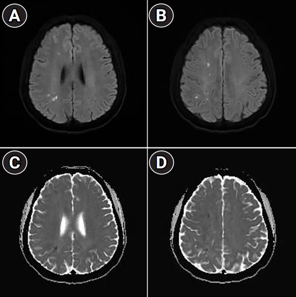
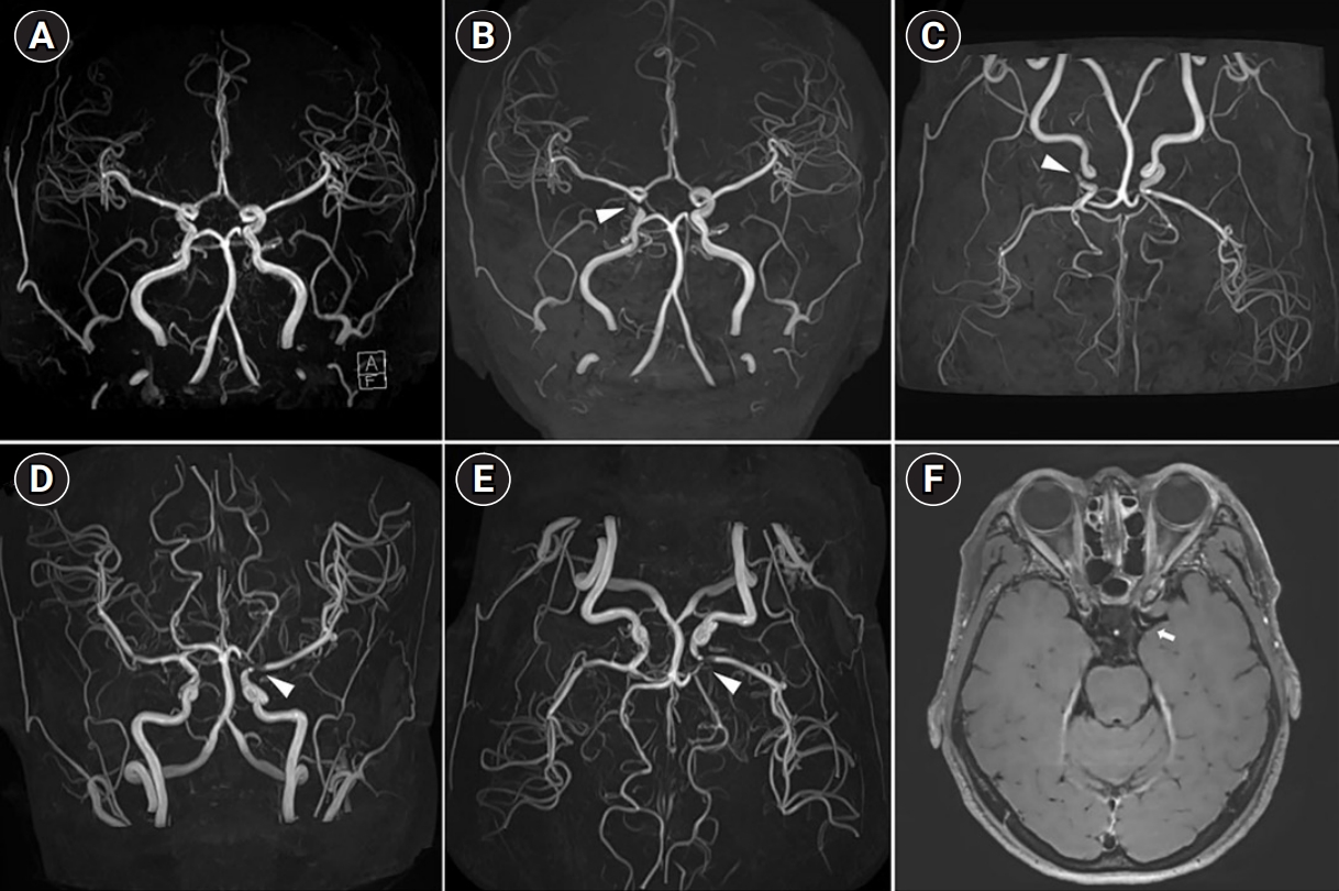
Extraglandular involvement in Sjögren's syndrome (SS) can manifest in the central or peripheral nervous system. Changes in major cerebral vessels in SS are rarely reported [1,2]. Pathomechanisms in SS are known to be mainly associated with small- or medium-sized vasculopathy [1,3]. A 59-year-old woman who presented with left transient hemiparesis a few days prior was diagnosed with SS with dry mouth. Anti-SS-related antigen A (SSA/Ro) and B (SSB/La) had shown strong positivity on previous evaluations. Brain magnetic resonance imaging (MRI) showed multiple diffusion-restricted lesions in the right middle cerebral artery border zone area, suggesting acute infarctions (Fig. 1). Magnetic resonance angiography (MRA) upon admission showed moderate-to-severe stenosis of the right distal internal carotid artery (ICA) (Fig. 2). The patient reported transient right hemi hypesthesia following admission. On MR carotid plaque imaging performed 4 days later, there was focal concentric wall thickening with enhancement in the right distal ICA stenosis at the location of the lesion shown on the previous MRA. Additionally, concentric wall thickening with enhancement was observed in the left distal ICA (Fig. 2). Bilateral distal ICA stenosis with wall thickening and enhancement occurred consecutively. Therefore, it was likely caused by vasculitic stenosis associated with SS.
This report showed that a patient with high disease activity developed severe vasculitic stenosis that occurred consecutively within a few days. A high-resolution MRI of the carotid plaque indicated the presence of vasculitis with large-artery involvement, which could be helpful when considering the pathology.

ARTICLE INFORMATION

Ethics statement
This work was approved by the Ethics Committees of the Inje University Busan Paik Hospital (No. 2015-01-271), and written informed consent was obtained from the patient.
Conflict of interest
No potential conflict of interest relevant to this article.
Acknowledgments
This study was supported by the National Research Foundation of Korea (NRF) grant funded by the Korea government (MIST) (No. 2020R1G1A1008446).
Author contributions
Conceptualization: WJK, SIO. Methodology: WJK, SIO. Formal analysis: WJK, SIO. Data curation: all authors. Visualization: all authors. Project administration: SIO. Writing–original draft: all authors. Writing–review and editing: WJK, SIO.

Fig. 1.
Brain magnetic resonance imaging scans upon admission. (A, B) There were high signal-intensity lesions on diffusion-weighted images in the right middle cerebral artery borderzone area at the right frontal and parietal lobes, and (C, D) relevant lesions were noted on the apparent diffusion coefficient map.
jnc-220088f1.jpg
Fig. 2.
Brain magnetic resonance angiography (MRA) and high-resolution magnetic resonance imaging (MRI) of the carotid plaque. (A) MRA taken 1 year before admission showed no stenosis in both distal internal carotid arteries (ICAs). (B, C) MRA taken upon admission showed moderate to severe stenosis in the right distal ICA (arrowhead). (D, E) On MRA taken 4 days later, severe stenosis was newly discovered in the left distal ICA, proximal M1, and proximal A1 (arrowhead), and (F) focal concentric wall thickening with enhancement at the left distal ICA was confirmed on high-resolution MRI (arrow).
jnc-220088f2.jpg

REFERENCES

1. Fox RI. Sjögren's syndrome. Lancet 2005;366:321-31.
crossref pmid
2. Tobón GJ, Pers JO, Devauchelle-Pensec V, Youinou P. Neurological disorders in primary Sjögren's syndrome. Autoimmune Dis 2012;2012:645967.
crossref pmid pmc
3. Li JA, Meng HM, Cui ZT, Wang X, Miao J. Recurrent cerebral infarctions in primary Sjögren syndrome: a case report and literature review. Front Neurol 2018;9:865.
crossref pmid pmc
Editorial Office
Department of Neurology and Critical Care, Seoul National University Hospital,
101, Daehak-ro, Jongno-gu, Seoul, 03080 Korea
TEL: +82-2-2072-0743 (10:00 AM ~ 4:00 PM) / +82-2-2072-1810   FAX: +82-2-3672-7553    E-mail: office@e-jnc.org

Copyright© Korean Neurocritical Care Society.                Developed in M2PI