INTRODUCTION
Patients in neuroscience intensive care unit (NSICU) frequently require invasive mechanical ventilation for concomitant respiratory failure. Intensivists need to pay special consideration to patients with elevated intracranial pressure (ICP) on invasive mechanical ventilation, particularly to ensure the maintenance of normal brain tissue oxygenation and optimal partial pressure of carbon dioxide (PCO2) levels, and to provide sedation that achieves both comfort and optimal patient-ventilator synchrony. We report a case of a life-threatening complication of refractory ICP elevation and a well-recognized complication of invasive mechanical ventilation—pneumomediastinum. The pathophysiological mechanisms and proposed treatment strategies are discussed.
CASE REPORT
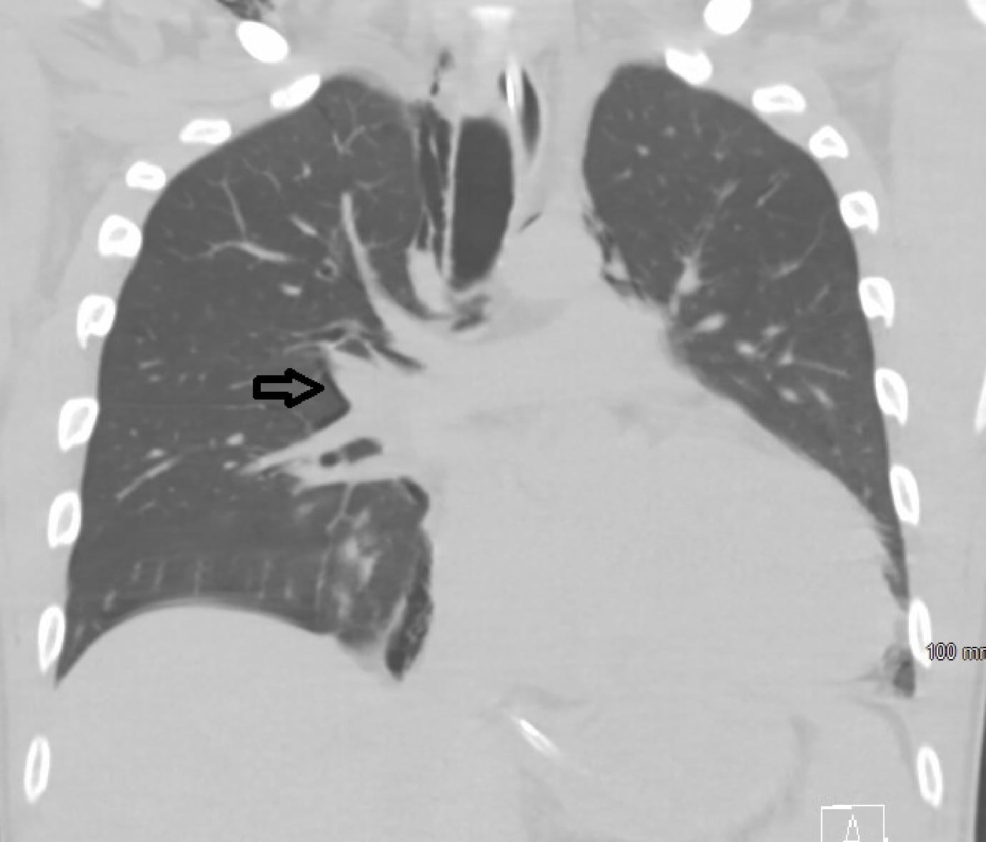
A 26-year-old man with a distant history of chronic myelogenous leukemia presented to the emergency department with headache, nausea, and vomiting. Computed tomography of the head revealed bilateral cerebellar lesions with surrounding vasogenic edema, resulting in a mass effect on the fourth ventricle and early tonsillar herniation. Subsequent biopsy was consistent with myeloid sarcoma, and the patient was started on steroids and whole-brain radiation. An external ventricular drain was placed due to progressive obstructive hydrocephalus on hospital day 7, but with progressive elevation of ICP, drowsiness, and poor tolerance of airway secretions, he was intubated and underwent suboccipital craniectomy on hospital day 12, with subsequent improvement of his ICP and associated symptoms. Postoperatively, the patient remained drowsy and struggled with impaired bulbar function and an inability to clear secretions, requiring continuous neurologic monitoring and prohibiting planned therapy with intrathecal chemotherapy and radiation. Although hemodynamically uncompromised, he ultimately needed reintubation due to respiratory fatigue on hospital day 14. Following an uncomplicated intubation, he was noted to have neck swelling and an increasing ICP trend. A computed tomography of the head and neck was ordered to rule out hematoma development in the setting of a recent suboccipital craniotomy (Fig. 1).
However, a post-intubation chest radiograph and subsequent examination revealed the presence of a new pneumomediastinum and subcutaneous emphysema (SQE) (Figs. 2 and 3). The patient continued to deteriorate clinically with rising ICP despite deep sedation. He had declining oxygen saturation despite a new 80% fraction of inspired oxygen (FiO2) requirement and the application of 6 cmH2O of positive end-expiratory pressure. He also had been developing hypotension with intermittent electrical alternans that required the initiation of vasopressors. Tension physiology impairing the intracranial venous drainage was suspected. An emergent tracheostomy was performed to relieve the patient’s tension pneumomediastinum while anticipating prolonged intubation, which might otherwise have limited his access to definitive therapy. Standard tracheostomy was performed midway between the sternal notch and thyroid cartilage at the level of the second tracheal ring, and allowed to heal by secondary intention to allow air to dissect across planes and relieve tension. A tracheostomy was successfully performed, resulting in decreased neck swelling, SQE, and pneumomediastinum. His ICP was noted to immediately decrease to 12–15 mmHg at the time of tracheostomy completion and remained stable for the following days; vasopressors were discontinued, and his oxygen requirements resolved. A limited proximal airway survey at the level of the segmental bronchi revealed no notable tracheal injury. The patient tolerated the transfer out of the NSICU for radiation therapy two days later. Ultimately, he required a ventriculoperitoneal shunt and was decannulated prior to being discharged home after a 103-day length of stay.
DISCUSSION
Successful management of intracranial hypertension is crucial to mitigate morbidity and mortality in neurocritical care. The tiered approach recommended for treatment starts with tier 0, which includes the management of pain, fever, anxiety, and proper positioning of the patient. Neutral positioning of the neck and elevation of the head of the bed to 30°; the proposed mechanism is the maximization of cerebral venous outflow and subsequent lowering of ICP [1]. Our case demonstrates how proper assessment of the cardiovascular and respiratory systems and early recognition of this rare complication can lead to improvements in our approach to lower ICP.
Intracranial pathology leading to acute respiratory failure may, in turn, intensify detrimental effects on the acutely injured brain. Respiratory failure, whether secondary acute hypoxic, hypercapnic, or ineffective ventilation due to inability to protect the airway from secretions or upper airway obstruction, is common in the NSICU [2]. The cilia of the nasopharynx function to propel secretions in a caudal direction, whereas those of the lower airways propel secretions in a cephalad direction. These secretions then accumulate in the pharynx to be swallowed or expectorated through complex airway protective mechanisms, which may be significantly impaired in neurological injury [3,4]. In the present case, posterior fossa pathology and direct brainstem compression leading to bulbar pathology presented a risk of acute aspiration and respiratory insufficiency. Increased work of breathing in patients with limited arousal and variable strength requires vigilance that may prohibit disposition from the intensive care unit or, as in this case, lead to intubation for airway protection.
Pneumomediastinum is a rare complication of intubation, although it was likely a higher risk in our patient due to the baseline anatomic abnormality of the pectus excavatum. Air accumulation, worsened by the application of positive pressure ventilation, led to rapid accumulation of mediastinal and subcutaneous air. Pneumomediastinum, with or without SQE, is a well-recognized complication of invasive mechanical ventilation. Alveoli may rupture due to different mechanisms, including alveolar hyperinflation and/or dynamic airway obstruction with increased intra-alveolar pressure. Air then escapes the alveolar space into the interstitium, dissecting its way from the peri-alveolar space following the bronchovascular structures to the hilum, and then accumulates in the mediastinum and up to the soft tissue of the neck, resulting in pneumomediastinum and concomitant SQE. This ascending pathway was described by Macklin [5] in 1939.
Pneumomediastinum is usually benign and well-tolerated by most patients. However, tension physiology has been described experimentally in the works of Macklin [5] through compression of the pulmonary veins, leading to increased afterload of the right ventricle. While pneumomediastinum does not typically cause rapid pressurization of the intrathoracic space, chest wall and parenchymal compliance may be restricted by the underlying anatomic or pathologic conditions. Patients may develop pneumopericardium and tension physiology that resembles tamponade [6,7]. If sufficient to cause hemodynamic compromise and shock, particularly obstructive physiology with evidence of electrical or pulsus alternans, narrowing of pulse pressures, and worsening hypoxia and/or hypotension, it follows that venous return from the intracerebral vasculature to the right heart will be limited [8]. In our case, the patient had progressive tension pneumomediastinum, likely in the setting of chronic restrictive lung disease; this clinically resulted in the direct compression of intrathoracic structures as well as vasculature in the neck, leading to a rapid rise in intracranial pressure and clinical deterioration.
Treatment of pneumomediastinum is usually achieved conservatively if not associated with hemodynamic compromise or other end-organ perfusion compromise (i.e., tension pneumomediastinum), and it may take one to two weeks for air to be fully absorbed in the absence of additional inspiratory pressures. On the other hand, tension pneumomediastinum can be fatal if not recognized and treated promptly. Similar to treating tension pneumothorax, rapid decompression of air to equilibrate the pressure gradient is desired. Surgical decompression (i.e., mediastinotomy) proposed by Macklin can be achieved through chest tube insertion in the subxiphoid region or through a simple suprasternal incision [5,8]. A simple incision might be more appealing in cases with no continuous air leak or in emergent conditions in which there is clear communication between the mediastinal air and suprasternal subcutaneous tissue.
Our patient developed refractory ICP elevation, hemodynamic compromise, and evidence of rapidly progressive obstructive shock, all of which resolved immediately after performing tracheostomy. Although higher than suprasternal mediastinotomy, pressurized air dissection through tissue planes along the path of least resistance to a tracheotomy site was sufficient to depressurize the neck and thorax, alleviating direct compression of the jugular veins and mediastinal structures, allowing right ventricular filling, and consequently, promoting cerebral venous drainage. We postulate that there is a critical pressure point beyond which further accumulation of air in the mediastinum leads to impaired venous drainage in the brain. In patients with refractory ICP elevation, we propose to routinely assess for the presence of SQE and pneumomediastinum. To our knowledge this is the first case of an adult with refractory ICP elevation due to pneumomediastinum. Furthermore, decompression of the pneumomediastinum is not often necessary, and tracheostomy may be considered in the rare event when intervention is required.