Posterior reversible encephalopathy syndrome as delayed neurological sequelae after carbon monoxide intoxication
Article information

A 27-year-old woman attempted suicide and was diagnosed with acute carbon monoxide (CO) poisoning. Neurological examinations were normal except for confusion. Initial brain images showed no acute lesions (Fig. 1A) on the next day of admission. Twelve days after CO intoxication, severe headache, choreic movement of both upper extremities, and impaired visual acuity with optic ataxia, oculomotor apraxia, and simultanagnosia were observed. Cerebrospinal fluid findings showed pleocytosis (white blood cell, 135/mm3) with elevated protein level (134.8 mg/dL). From the follow-up brain magnetic resonance imaging, she was diagnosed with posterior reversible encephalopathy syndrome (PRES) as delayed neurologic sequelae (DNS) after CO intoxication (Fig. 1B). With steroid pulse therapy, she had clinical improvement (Fig. 1C). During the acute phase of CO poisoning, brain MRI shows signal changes in the bilateral globus pallidus (GP) with cytotoxic edema [1]. PRES shows distinctive MRI findings of the parieto-occipital lesions with either vasogenic or cytotoxic edema or both [2]. A patient with DNS with PRES was reported without GP involvement [3]. However, in this case, DNS after CO intoxication affected not only the basal ganglia but also the parieto-occipital regions as PRES. Therefore, it is the reason that the patient presented with both chorea and Balint’s syndrome simultaneously.
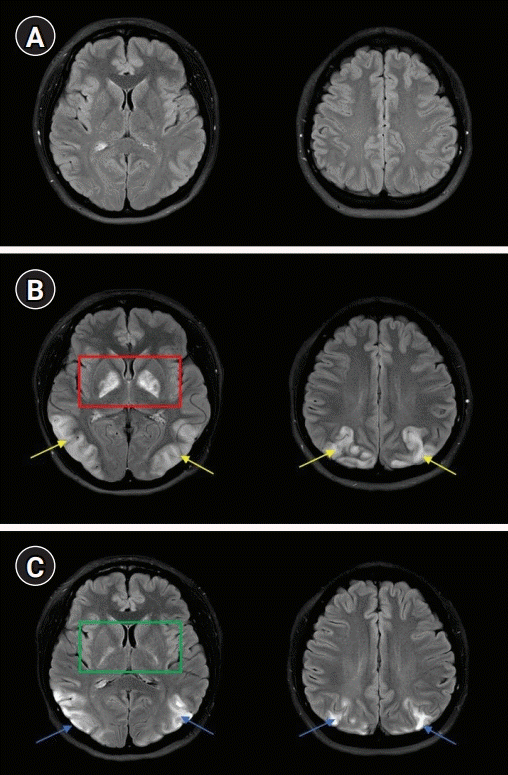
Brain magnetic resonance imaging. (A) The day of carbon monoxide (CO) poisoning, fluid-attenuated inversion recovery (FLAIR) images showed no abnormal high signal intensity. (B) Twelve days from CO intoxication, FLAIR images showed high signal intensities in bilateral globus pallidus (red box) and both parieto-occipital cortex (yellow arrows), suggesting posterior reversible encephalopathy syndrome. (C) Twenty-five days after CO intoxication, follow-up FLAIR image was obtained from the outpatient visit, and it showed interval decrease of signal and volume in bilateral globus pallidus (green box) and temporo-parieto-occipital cortex (blue arrows).
Notes
Ethics statement
This case was reviewed and approved by the Institutional Review Board of Hanyang University Hospital (IRB No. 2021-10-012). The need for informed consent from a patient was waived by the IRB.
Conflict of interest
No potential conflict of interest relevant to this article.
Funding
This research was supported by the research fund of Hanyang University (HY-202100000001136).
Author contributions
Conceptualization: JP. Data curation: all authors. Formal analysis: all authors. Funding acquisition: JP. Investigation: all authors. Project administration: all authors. Visualization: all authors. Writing–original draft: all authors. Writing–review & editing: all authors.