Chung, Lee, and Kim: Serial brain magnetic resonance imaging in patient with invasive streptococcal infection with ventriculitis and choroid plexitis
A 39-year-old previously healthy man visited the hospital with fever, myalgia, and vomiting. His systolic blood pressure was below 60 mmHg, and he was admitted to the intensive care unit with the suspicion of septic shock. After admission, Streptococcus pneumonia was found in his blood, and treatment with vancomycin 2 g/day and ceftriaxone 4 g/day was initiated. Following antibiotic treatment for 4 days, his mentation deteriorated to a stupor. Brain magnetic resonance imaging (MRI) indicated hydrocephalus, ventriculitis, and choroid plexitis ( Fig. 1). Cerebrospinal fluid (CSF) examination showed normal opening pressure (180 mmHg), pleocytosis (white blood cell count, 280; neutrophils, 55%), high protein level (1,448 mg/dL), and low glucose levels (CSF, 54 mg/dL; serum, 135 mg/dL). Consequently, a high dose of steroids (dexamethasone, 40 mg/day) was prescribed together with antibiotics. After 14 days of antibiotic treatment, his general condition improved. Brain MRI findings depicting pyogenic ventriculitis typically include ependymal thickening and enhancement with T2 prolongation surrounding the ventricle, hydrocephalus, and debris within the dependent aspect of the ventricles [ 1, 2]. In addition, diffusion restriction and swelling of the choroid plexus are suggestive of choroid plexitis [ 2, 3]. Although the MRI findings of our patient seemed more critical than previous reports, the patient experienced a good outcome with antibiotics [ 4].
Fig.┬Ā1.
Serial brain magnetic resonance imaging (MRI) findings of invasive streptococcal sepsis with ventriculitis and choroid plexitis. Initial brain images reveal hydrocephalus with severe interstitial edema and non-suppressed high-signal fluid in the lateral ventricle on fluid-attenuated inversion recovery (FLAIR) imaging, suggesting pyogenic ventriculitis. In addition, restricted diffusion on diffusion-weighted imaging (DWI) and low signal intensity on susceptibility-weighted imaging (SWI) in both choroid plexuses indicate choroid plexitis with hemorrhaging (A). Although the patientŌĆÖs mental status improved after administration of intravenous vancomycin and ceftriaxone, no significant change is observed in the brain MRI on day 7 after admission (B). The brain MRI of day 45 shows improved hydrocephalus, interstitial edema, ventriculitis, and choroid plexitis; however, a high signal of cerebrospinal fluid on FLAIR imaging is noted (C). Finally, on day 300, a normal brain MRI is observed (D). 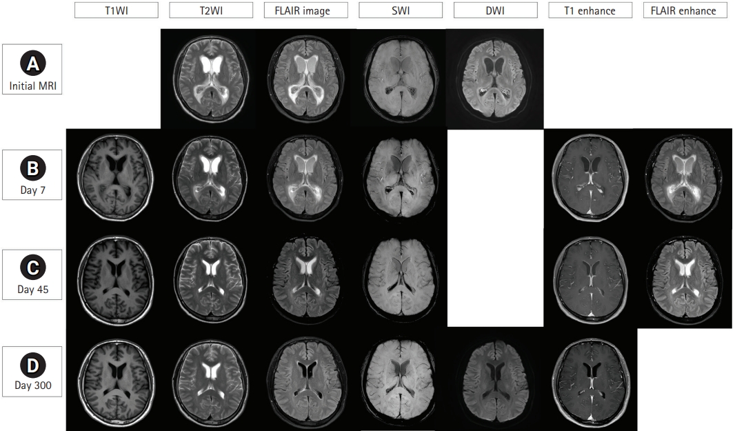
REFERENCES
1. Mohan S, Jain KK, Arabi M, Shah GV. Imaging of meningitis and ventriculitis. Neuroimaging Clin N Am 2012;22:557-83.   2. Hazany S, Go JL, Law M. Magnetic resonance imaging of infectious meningitis and ventriculitis in adults. Top Magn Reson Imaging 2014;23:315-25. 3. Pezzullo JA, Tung GA, Mudigonda S, Rogg JM. Diffusion-weighted MR imaging of pyogenic ventriculitis. AJR Am J Roentgenol 2003;180:71-5. 4. Woehrl B, Linn J, Lummel N, Pfefferkorn T, Koedel U, Pfister HW, et al. Pneumococcal meningitis-associated pyogenic ventriculitis. J Infect 2015;70:311-4.
|
|


