Which one to do first?: a case report of simultaneous acute ischemic stroke and myocardial infarction
Article information

Abstract
Background
Although there are common risk factors for acute ischemic stroke and myocardial infarction, simultaneous onset of both diseases is uncommon. Here, we present a case of acute cerebral infarction with concurrent fatal myocardial infarction.
Case Report
A 54-year-old man presented with left hemiparesis, gaze preponderance to the right side, and visual and tactile extinction. Computed tomography angiography showed occlusion of the right middle cerebral artery. ST-elevation myocardial infarction was suspected on electrocardiography. After the injection of intravenous tissue plasminogen activator, thrombectomy was attempted first, and the coronary angiogram was planned after recanalization of the cerebral artery. However, thrombectomy was discontinued because of cardiac arrest. Despite extracorporeal membrane oxygenation and emergency percutaneous coronary intervention, the patient died of multiorgan failure.
Conclusion
Double primary acute ischemic stroke and myocardial infarction are rare but may be fatal due to the narrow therapeutic time window for both diseases. Careful consideration of the urgency of cardiac status is essential.
INTRODUCTION
Acute ischemic stroke (AIS) and acute myocardial infarction (AMI) are life-threatening conditions that may lead to permanent morbidity or disability. Although there are many shared risk factors [1-3], simultaneous onset of both AIS and AMI is uncommon. In such rare circumstances, physicians are left with a dilemma of treating one condition may delay the treatment of the other condition. Here, we present a case of acute cerebral infarction that occurred concurrently with fatal AMI at the same time.
CASE REPORT
A 54-year-old man presented to the emergency room with left hemiparesis. Neurologic examination further showed gaze preponderance to the right side, central type left facial palsy, and visual and tactile extinction. He did not show asomatognosia or anosognosia. The National Institutes of Health Stroke Scale score was 17. The neurological deficit was first detected by a witness who reported him to the police because he had been driving his car and scratching the median strip. He was a smoker and had been taking medications for hypertension and diabetes mellitus. Brain computed tomography (CT) showed a focal low density in the right insula, corona radiata, and temporal lobe (Fig. 1A), and CT angiography showed occlusion of the M1 segment of the right middle cerebral artery (Fig. 1B). In the CT perfusion image, the Tmax value was increased in the right middle cerebral artery and posterior cerebral artery territory due to the fetal posterior cerebral artery (Fig. 1C). Because he was alert and denied having any neurological symptoms when he started driving at 16:00, intravenous tissue plasminogen activator (tPA) was injected at 18:35. The door-to-needle time was 59 minutes. Initial electrocardiogram showed ST-segment elevation in leads II, III, and aVF with reciprocal ST depression in V5 and V6, suggesting acute inferior myocardial infarction (Fig. 2A). Although initial creatinine kinase (CK) and CK-myocardial band were within the normal range, troponin-I was elevated to 0.130 ng/mL and N-terminal-pro hormone B-type natriuretic peptide was elevated to 1,859 pg/mL. Endovascular thrombectomy (EVT) of the thrombus in the right middle cerebral artery was attempted first, planning the coronary angiogram after the recanalization of the cerebral artery, since the patient was alert and did not report any chest pain. However, thrombectomy was stopped without recanalization due to cardiac arrest during the procedure. Electrocardiography revealed a pulseless ventricular tachycardia. After cardiopulmonary resuscitation for 21 minutes, extracorporeal membrane oxygenation was performed. An emergency coronary angiogram showed a culprit lesion in the right coronary artery (Fig. 2B), with other coronary arteries remaining intact. Percutaneous coronary intervention (PCI) was performed for the occluded right coronary artery to achieve recanalization of the right coronary artery (Fig. 2C). Despite intensive medical care for 5 hours with inotropic agents, the patient died after gradual blood pressure drop and progression of multi-organ failure.

(A) Brain computed tomography without enhancement demonstrates low density in the right insula, corona radiate, and temporal lobe (white arrowheads). Sulcal effacement is also noted in right frontal and temporal lobe. (B) Brain computed tomography angiogram demonstrates occlusion of the M1 segment of the right middle cerebral artery (white arrow). Severe stenosis is also noted in the right distal internal carotid artery (black arrowhead). (C) Perfusion image shows increased Tmax value in the right middle and posterior cerebral artery territory.
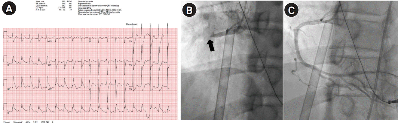
(A) Electrocardiogram shows ST-segment elevation in II, III, and aVF with reciprocal ST depression in V5 and V6, suggesting acute inferior myocardial infarction. (B) Emergency coronary angiogram shows the culprit lesion in the right coronary artery before stenting (black arrow). (C) After percutaneous coronary intervention, recanalization of right coronary artery is achieved.
DISCUSSION
Concurrent AIS and AMI, in other words, a cardiocerebral ischemic (CCI) attack, is an infrequent but a challenging medical condition [4,5]. The mechanism of this life-threatening situation is unclear and may be multifactorial, from the increased risk of intracardiac thrombus formation due to left ventricular systolic dysfunction, to catecholamine-induced myocardial stunning due to the adrenergic surge associated with cerebral infarction, especially in the insular cortex [6,7]. Moreover, there have been several case reports of CCI attacks in special circumstances, such as aortic dissection, electrical injury, marijuana abuse, or underlying acute myeloid leukemia [8-10]. In some cases, a CCI attack may also occur due to atherosclerosis in the cerebral and coronary arteries, respectively [11].
Regardless of the mechanism, a CCI attack may be fatal because the treatment of both AIS and AMI has a narrow therapeutic time window. The acute treatment of one condition can result in a critical delay in the other treatment. Although intravenous alteplase injection may be helpful for both cerebral and myocardial infarction, intra-arterial reperfusion therapy for the cerebral artery prior to PCI can cause a delay in PCI, even leading to cardiac arrest, as in the present case. Meanwhile, PCI prior to cerebral reperfusion therapy can increase the risk of a large cerebral infarction, which can cause severe neurological deficits, cerebral edema, and even death. Moreover, the use of antiplatelets, which are essential after PCI, may be harmful when the cerebral infarction is too large, with a substantial risk of hemorrhagic transformation [5,11].
Despite such important clinical needs, there are no evidence-based guidelines or clinical studies on the management of the co-occurrence of AIS and AMI, especially in the priority of the treatment [7]. Most of the recent studies on CCI are case reports or case series describing various cerebral and myocardial infarct territories, heterogeneous timing and modalities of treatment, and consequent various outcomes [12,13]. Omar et al. [5] reported the case of a 48-year-old patient with inferior-posterior and right ventricular AMI concomitant vertebra-basilar territory AIS, who was treated with tPA and conservative management, and expired on the second day of hospital stay. In contrast, Yeo et al. [4] reported the case of a 53-year-old patient who was treated with PCI and stenting first, followed by EVT. The patient survived with aphasia and disability that required a wheelchair. In such case reports, the choice of treatment varies from conservative management with antithrombotics to aggressive management, such as PCI and EVT.
Because PCI and EVT are becoming more available these days, the choice of treatment for CCI is becoming more complex. A single-center case series reported that among nine patients who presented with synchronous onset of AMI and AIS, one patient underwent PCI, another patient underwent intravenous thrombolysis, and the others only received conservative management, leaving six survivors [13]. Meanwhile, in a meta-analysis of case reports and series describing the patient characteristics, investigations demonstrated, treatments, and outcomes [14], 10 out of the 44 enrolled patients died within a median of 2 days, despite the aggressive treatment of PCI with stenting in 15 patients, PCI without stenting in eight patients, thrombectomy of a coronary vessel in eight patients, and EVT in 10 patients. Even with emergent and aggressive treatments, the prognosis of CCI is still devastating. Moreover, the optimal order of PCI and EVT remains unclear.
Guidelines for the early management of patients with acute ischemic stroke, updated in 2019, recommend intravenous alteplase at the dose used for cerebral ischemia followed by PCI for hyperacute co-occurrence of AIS and AMI (class IIa; level of evidence C) [15]. However, the dosage needed for the management of AMI differs from that used in the treatment of AIS [12,13]. Moreover, tPA can increase the risk of cardiac wall rupture or tamponade in patients with AMI. Due to the lack of optimal treatment guidelines, careful consideration of the urgency of the cardiac status and tailored treatment strategies are essential in patients with simultaneous AIS and AMI. According to a case series report, most CCI patients (83%) presented only with neurological deficits without chest pain [13], as in our case. A high level of suspicion is necessary to improve the recognition and management of patients with CCI. Emergency bedside echocardiography can be helpful in evaluating cardiac function and deciding the order of treatment. Further large observational studies and randomized trials are need to be conducted in order to recommend the optimal treatment for this subset of patients.
Notes
Ethics statement
In accordance with the principles of the Institutional Review Board (IRB) of National Health Insurance Service Ilsan Hospital that a case report of three or less cases does not require IRB approval, the need for IRB approval and informed consent from a patient was waived.
Conflict of interest
No potential conflict of interest relevant to this article.
Author contributions
Conceptualization: LKJ. Data curation: PWH. Formal analysis: LKJ, SKD. Methodology: LKJ, KHS, SKD. Project administration: LKJ. Visualization: LKJ, KHS. Writing–original draft: LKJ. Writing–review & editing: SKD.