| Home | E-Submission | Sitemap | Contact Us |  
Journal of Neurocritical Care > Volume 12(1); 2019 > Article
Kim and Jeon: Cerebral air embolism treated using hyperbaric oxygen therapy
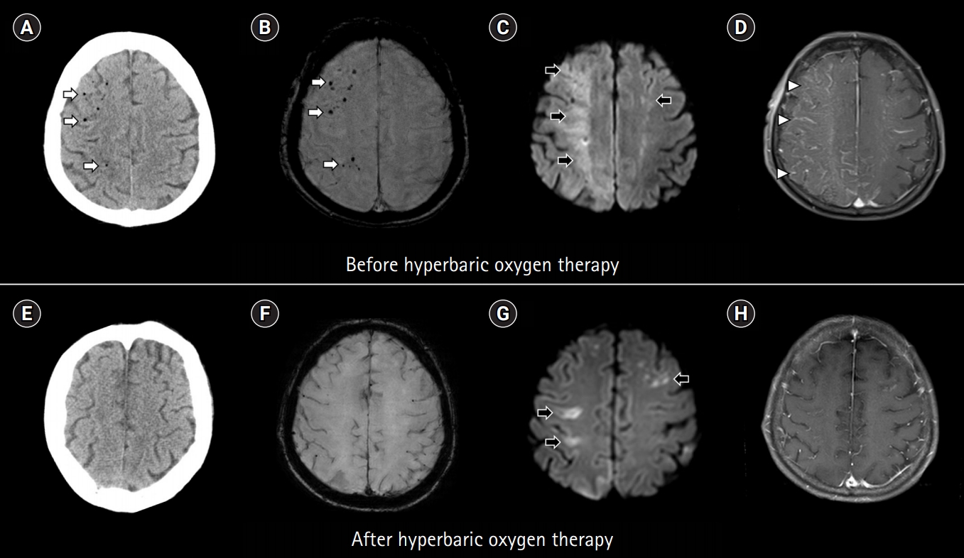
A 59-year-old man underwent endoscopic balloon dilation for esophageal stricture. Upon the completion of the procedure, he was observed to be stuporous and quadriplegic. Computed tomography (CT) of the brain revealed multiple air emboli (Fig. 1A). Magnetic resonance imaging (MRI) performed 45 minutes after symptom onset revealed multiple lesions showing signal loss on susceptibility-weighted imaging, high signal intensity on diffusion-weighted imaging, and diffuse enhancement on T1-weighted contrast-enhanced imaging, which were compatible with air emboli, hyperacute infarcts, and a disrupted blood-brain barrier, respectively (Fig. 1B-1D). Hyperbaric oxygen therapy (HBOT) was administered 80 minutes after MRI (targeting 3.0 atmospheric pressure for 2 hours). Follow-up CT performed 80 minutes after HBOT revealed disappearance of the air emboli (Fig. 1E). Follow-up MRI performed 5 days after HBOT also revealed a decrease in the resolution of previously documented findings (Fig. 1F-1H). Most of his neurological symptoms improved, except mild left hemiparesis.
This case indicates that MRI is a useful modality in diagnosing cerebral air embolism by documenting air emboli, hyperacute infarcts, and disruption of the blood–brain barrier [1-3]. HBOT may accelerate the disappearance of air emboli and promote the early resolution of ischemic lesions and reversal of the disrupted blood–brain barrier [4].

ARTICLE INFORMATION

Conflict of interest
Dr. SB Jeon is an editorial board member of the journal but was not involved in the peer reviewer selection, evaluation, or decision process of this article. There are no other potential conflicts of interest relevant to this article to declare.
Funding
This research was supported by a grant of the Korea Health Technology R&D Project through the Korea Health Industry Development Institute (KHIDI), funded by the Ministry of Health & Welfare, Republic of Korea (grant number : HI18C1487).
Author contributions
Conceptualization: YJK and SBJ. Data curation & Formal analysis: YJK and SBJ. Visualization & Writing–original draft: YJK and SBJ. Writing–review editing: YJK and SBJ.

Fig. 1.
Computed tomography (CT) and magnetic resonance imaging (MRI) scans of the brain obtained before (upper row) and after (lower row) hyperbaric oxygen therapy (HBOT). (A) Initial CT scan showing multiple air emboli, predominantly in the right hemisphere. (B) Initial MRI scan showing multiple air emboli on susceptibility-weighted imaging, (C) acute infarcts on diffusion-weighted imaging, and (D) diffuse enhancement on T1-weighted contrast-enhanced imaging. The Glasgow Coma Scale score was 5 (eye opening, 2; motor response, 2; and verbal response, 1) when the initial MRI scan was obtained. (E) Follow-up CT scan obtained 80 minutes after HBOT showing disappearance of air emboli. (F) Follow-up MRI scan obtained 5 days after HBOT showing disappearance of air emboli on susceptibility-weighted imaging, (G) decrease in infarct size on diffusion-weighted imaging, and (H) resolution of diffuse enhancement on T1-weighted contrast-enhanced imaging. The Glasgow Coma Scale score was 14 (eye opening: 4, motor response: 6, and verbal response: 4) when the follow-up MRI scan was obtained. White arrows indicate air emboli, black arrows indicate acute infarcts, and white arrowheads indicate diffuse T1-enhancement.
jnc-190085f1.jpg

REFERENCES

1. Jeon SB, Kim JS, Lee DK, Kang DW, Kwon SU. Clinicoradiological characteristics of cerebral air embolism. Cerebrovasc Dis 2007;23:459-62.
crossref pmid
2. Jeon SB, Kang DW. Cerebral air emboli on T2-weighted gradient-echo magnetic resonance imaging. J Neurol Neurosurg Psychiatry 2007;78:871.
crossref pmid pmc
3. Hjouj M, Last D, Guez D, Daniels D, Sharabi S, Lavee J, et al. MRI study on reversible and irreversible electroporation induced blood brain barrier disruption. PLoS One 2012;7:e42817.
crossref pmid pmc
4. Gill AL, Bell CN. Hyperbaric oxygen: its uses, mechanisms of action and outcomes. QJM 2004;97:385-95.
crossref pmid pdf
Editorial Office
Department of Neurology and Critical Care, Seoul National University Hospital,
101, Daehak-ro, Jongno-gu, Seoul, 03080 Korea
TEL: +82-2-2072-0743 (10:00 AM ~ 4:00 PM) / +82-2-2072-1810   FAX: +82-2-3672-7553    E-mail: office@e-jnc.org

Copyright© Korean Neurocritical Care Society.                Developed in M2PI