Detection of neck hematoma after carotid endarterectomy by chest X-ray
Article information

A 61-year-old man presented to the emergency room with left hemiparesis and dysarthria. Brain magnetic resonance imaging showed right middle cerebral artery territory infarction with proximal internal carotid artery stenosis. Carotid endarterectomy (CEA) was performed on the 8th day of admission. After 3 hours of CEA, the patient complained of dyspnea, and stridor was developed. Chest X-ray was performed immediately (Fig. 1), and emergent endotracheal intubation was performed under suspicion of airway obstruction due to hematoma. Neck computed tomography (CT) confirmed hematoma formation around the carotid vessels (Fig. 2). Blood pressure was strictly controlled without discontinuation of clopidogrel, and hematoma was noted to be reduced on a CT scan 10 days after intubation. The patient was extubated without airway problems.

(A) Chest X-ray taken before carotid endarterectomy (CEA) shows normal findings. (B) Chest X-ray taken during dyspnea after CEA shows soft tissue swelling (black arrow) with tracheal deviation (white arrow). (C) Chest X-ray taken the day after intubation shows aggravation of soft tissue swelling (black arrow).
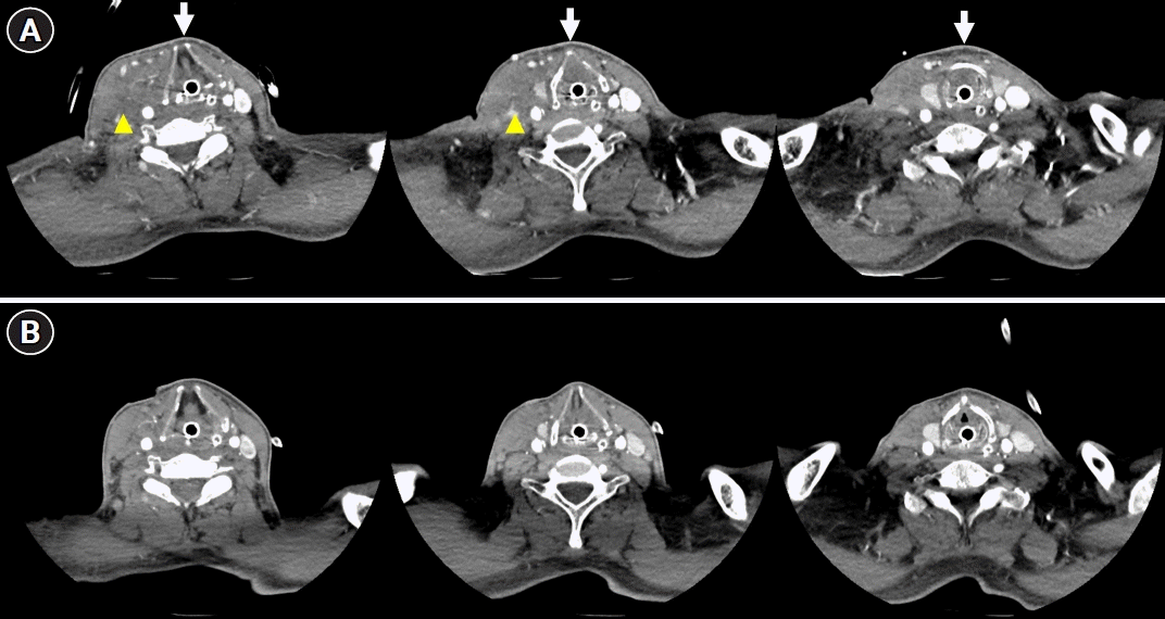
(A) Neck computed tomography (CT) taken after intubation shows tracheal deviation (white arrows) due to hematoma formation (yellow arrowheads) around the carotid vessels. (B) Neck CT taken 10 days later shows resorption of hematoma and improvement in tracheal deviation.
CEA is the standard treatment for symptomatic carotid stenosis [1,2]. A previous study reported that perioperative hematoma occurred in 7.1% of patients after CEA and was associated with increased perioperative stroke and mortality [3]. Neck hematoma is potentially life-threatening because it can cause respiratory failure and often requires airway management [2]. Suspicion of neck hematoma is crucial if the patient complains of respiratory discomfort after CEA, and airway management should be performed immediately [2]. Checking the chest X-ray can provide guidance in the differential diagnosis of respiratory discomfort after CEA.
Notes
Ethics statement
This study was reviewed and approved by the Institutional Review Board of Dong-A University Hospital (No. DAUHIRB-23-159). The need for informed consent from the patient was waived by the board.
Conflict of interest
Jin-Heon Jeong is an editorial board member of the journal, but he was not involved in the peer reviewer selection, evaluation, or decision process of this article. No other potential conflict of interest relevant to this article was reported.
Author contributions
Conceptualization: JJH. Methodology: all authors. Formal analysis: all authors. Data curation: all authors. Visualization: all authors. Project administration: all authors. Writing–original draft: all authors. Writing–review & editing: all authors.